

If you find this useful, share it with a friend, become a paid subscriber, or gift a subscription. Paid subscriptions give you full access to every article and help this Substack climb the rankings to reach more readers.
“This is truly groundbreaking” —Greg Harrison
“We had to find a lab, we had to fund it ourselves. It’s all out of our own pocket…
“We have forensic grade proof that the material is amyloid like… The proteins have not merely lost their native fold, they have adopted a new thermodynamically stable architecture.
“Data is data and facts are facts. And these are facts. So all we can do is present the facts…
“We challenge people. If you disagree with it, you go and rerun the work, you go and get the Raman analysis and prove us wrong.”
Greg Harrison is an organic chemist based in Sydney, Australia. For the past two years, he has been working with a small international team of scientists analyzing unusual white, rubbery, fibrous clots found inside the blood vessels of deceased people — and also in living people.
These white clots began appearing in corpses in late spring 2021, shortly after the mRNA vaccine rollout began, and they are still being found today. Embalmers were the first to notice them; vascular surgeons have since found them in living patients as well.
What Are These Rubbery White Structures or Clots?
Unlike normal red blood clots, they are not made of standard fibrin; the body cannot dissolve them through its normal clot-clearing processes. They are rubbery, rigid, and highly structured and resemble a scaffold or lattice, not the smooth spaghetti-like appearance of normal fibrin. Under 5000x microscopy, the structures appear twisted and abnormal.
While normal blood clots can be broken down naturally by the body via plasmin digestion, our body is unable to dissolve the white clots.
The Team’s Core Finding: Blood-Borne Systemic Amyloidosis
Greg’s team now believes these white clots are amyloid or amyloid-like which means that they consist of misfolded proteins that have locked into a rigid, crystalline structure.
Amyloidosis is a group of diseases that are caused by misfolded proteins building up in organs, including the brain. Alzheimer’s is one of them, Lewy body diseases another.
Greg told me that there are 36 known different types of amyloidosis in total and that the white clots are a completely new type of amyloidosis: a blood-borne systemic amyloidosis, never seen before.
Traditional amyloidosis develops slowly over decades. But this white clot amyloidosis develops much faster, namely within months. These white structures were seen by embalmers in corpses for the first time in spring of 2021, just months after the mRNA Covid mass injection campaign had begun.
Below you see one of Greg’s slides where he shows the Australian Amyloidosis Network’s definition:
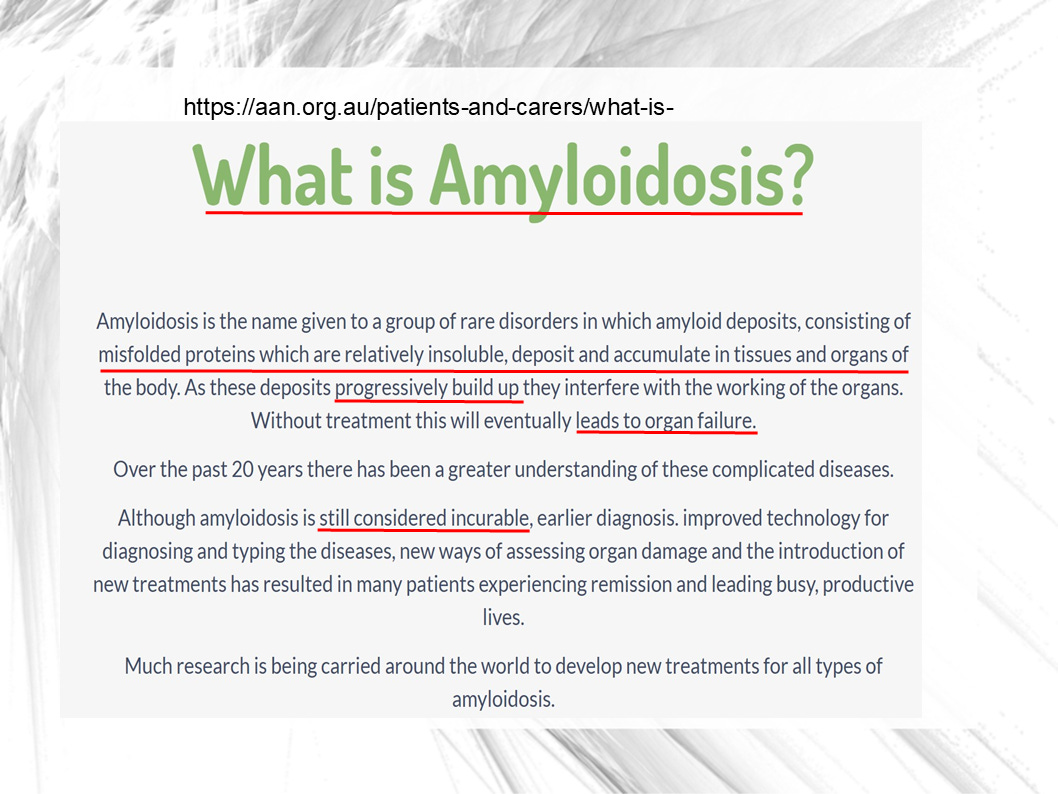
https://aan.org.au/patients-and-carers/what-is-amyloidosis/
The Spike Protein Effect: A New Type of Amyloidosis Caused by the Spike Protein
The man-made toxic spike protein, together with lipid nanoparticles, causes white clot amyloidosis. It triggers severe misfolding of the blood-clotting fibrinogen protein.
Plenty of fibrinogen lives in our blood and, when we suffer an open wound, converts into fibrin, a string like protein that clots the blood and prevents us from bleeding out.
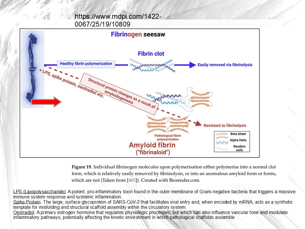
The slide below is from Greg Harrison’s Powerpoint presentation and shows the difference between a healthy fibrin clot formation and the pathological amyloid fibrin formation caused by the spike protein.
Our bodies have no problem removing fibrin clots via fibrinolysis once the clot has done its job in stopping the bleeding from a wound.
However, we are incapable of removing amyloid fibrin (white clots) from our bloodstream:
But what is the exact mechanism of this Spike protein effect?
The spike protein from a Covid-19 infection is in a so-called “closed” conformation, which means that its amyloid-promoting sequences are buried and can’t easily gain access to fibrinogen.
However, the mRNA vaccine-derived spike differs from the Covid infection spike protein: The vaccine-derived spike protein is “locked open” so that the amyloid sequences are exposed and can directly react with the large amount of fibrinogen in our blood.
Microclots Found in Injected and in Uninjected People
Nonetheless, amyloid microclots have been found not only in injected people but also in uninjected people who had Covid. However, microclots in the uninjected form much more slowly than in the injected apparently.
There are many questions about the body’s ability to digest the microclots triggered by Covid-19 or by mRNA injection. So far, nobody knows whether the body can dissolve them at all or up to which size.
The Evidence: The Tests Harrison’s Team & Dr. McCairn Ran
Greg Harrison’s small team of international scientists (who for now must remain anonymous to protect their careers) as well as neuroscientist Dr. Kevin McCairn ran many lab tests, some of them highly sophisticated and expensive ones that require specialized lab equipment. All images below are from Greg Harrison’s Powerpoint slide show.
ICP-OES & ICP-MS (elemental analysis): This elemental analysis found abnormally high levels of phosphorus and sulfur in the clots — phosphorus levels of 4,400–4,900 parts per million, orders of magnitude higher than normal.
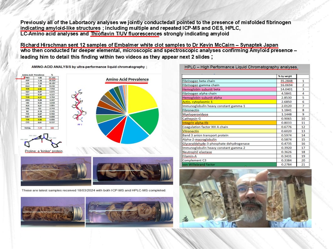
LCMS (liquid chromatography mass spectrometry): This test found proline as the most abundant protein in the clots — significant because the spike protein is locked open by two proline inserts. Proline is a unique protein-building amino acid (it’s really an imino acid). What’s important to know is that proline often shapes how protein folds: it causes tight turns and kinks in polypeptide chains and disrupts regular alpha helices and beta sheets. Which is exactly like the result of the misfolded fibrinogen.
HPLC (high performance liquid chromatography): HPLC found an unusually high concentration of fibrinogen, and the fibrinogen monomers were in an abnormal ratio.
On the image below, you see the results of this high performance liquid chromatography in the table on the right:
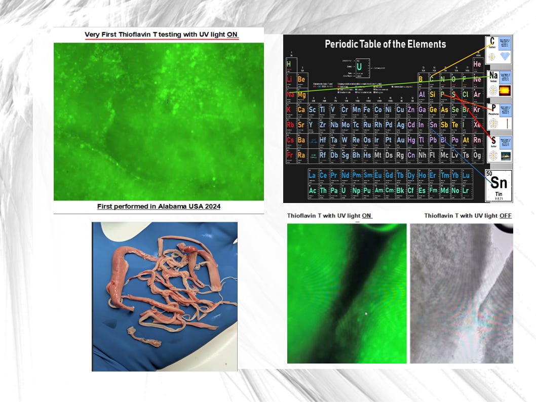
Thioflavin T staining (this is the gold standard amyloid test): The clots glowed bright green under UV light which is a direct indicator of the presence of amyloid structures; the brighter the glow, the more advanced the amyloid formation is.
As you can see, the green glows very brightly which means that a lot of amyloid structures are present in the sample.
Raman spectroscopy (this is the gold standard test for protein structure): This Raman spectroscopy was done at a world-renowned European laboratory by a recognized Raman spectroscopy expert; this test measures the exact vibrational energy of chemical bonds and can “fingerprint” protein structures.
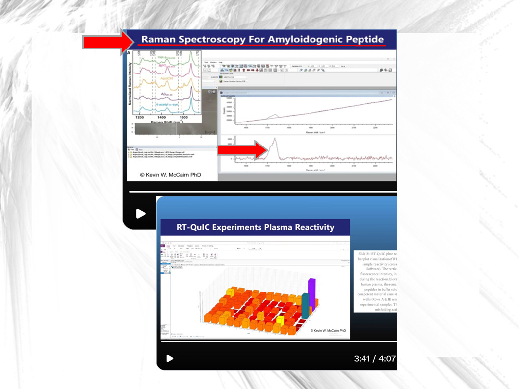
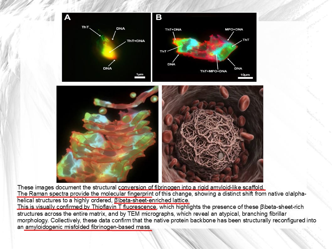
Results showed a transition from a flexible, normal alpha-helical protein structure to a beta-sheet enriched lattice — the hallmark of amyloid.
Below you see such an amyloid-like scaffold on the bottom right:
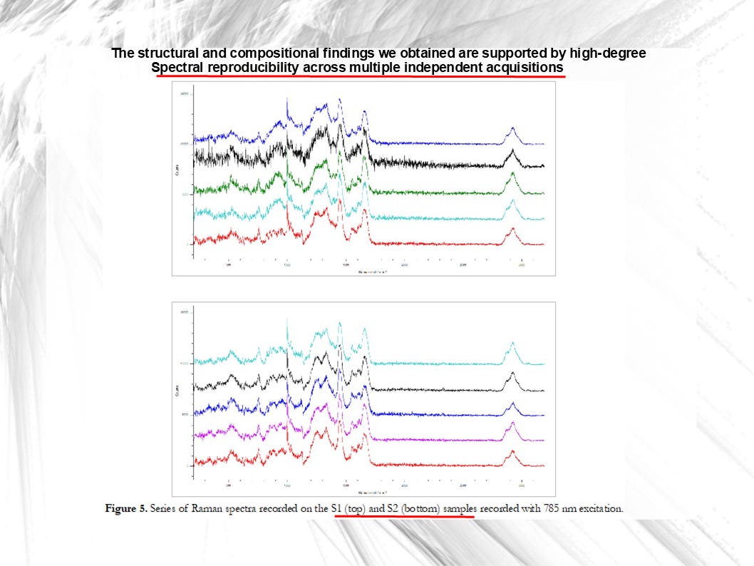
Five separate measurements taken from different parts of the same clot all gave a perfectly consistent result, see below:
A Raman fingerprint comparison confirmed the material matched misfolded fibrinogen, not normal fibrinogen.
The top graph compares Raman spectra of normal human blood plasma (upper trace) with purified crystalline human fibrinogen (lower trace) across wavenumber; the yellow-highlighted region marks the main band cluster used as a structural “fingerprint”.
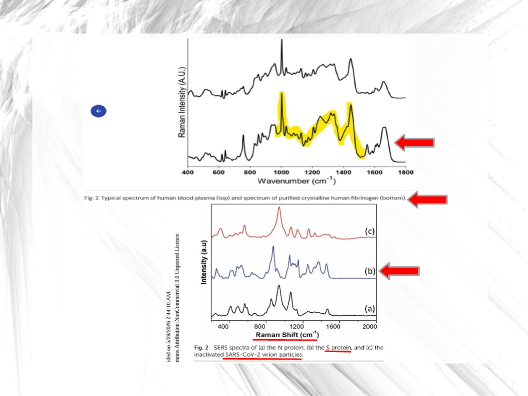
The bottom graph shows Raman spectra for three SARS‑CoV‑2 components: (a) the N protein, (b) the S protein, and (c) inactivated whole virion particles.
The N protein wraps and packages the viral RNA genome inside the virus particle, forming the ribonucleoprotein core.
The S protein or Spike protein forms the spikes on the virus surface and is the key molecule the virus uses to attach to and enter human cells via receptors like ACE2.
Below is a slide showing the actual Raman measurements of the clot samples.
Both graphs show two high‑resolution Raman fingerprints of clot samples, but measured at two different laser wavelengths (called “excitation”). The fact that they match each other so closely supports the claim that both specimens share the same underlying structured (and misfolded) protein lattice.
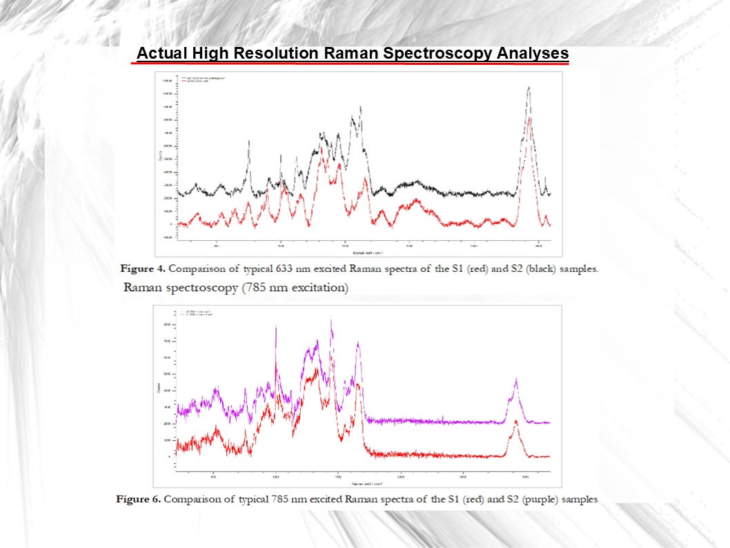
The top graph (Figure 4) compares Raman spectra taken at 633 nm excitation for two samples labeled S1 (red curve) and S2 (black curve) across wavenumber; you can see how their peaks line up and differ across the spectrum.
The bottom graph (Figure 6) does the same kind of comparison at 785 nm excitation, again plotting S1 (red) against S2 (purple) to show that the spectral “fingerprints” of both samples are very similar over the Raman shift range.
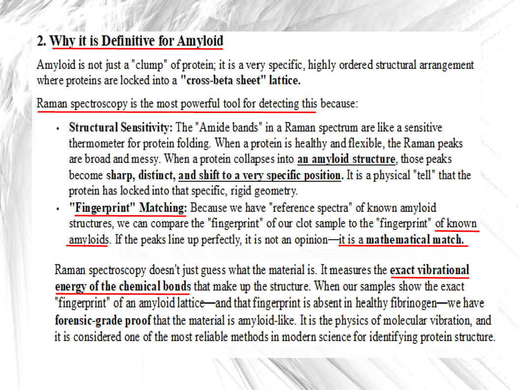
On the slide below, Greg Harrison explains why Raman spectroscopy is the most powerful and most reliable tool for detecting amyloid - proteins misfolded that become locked into a cross-beta sheet lattice-like structure:
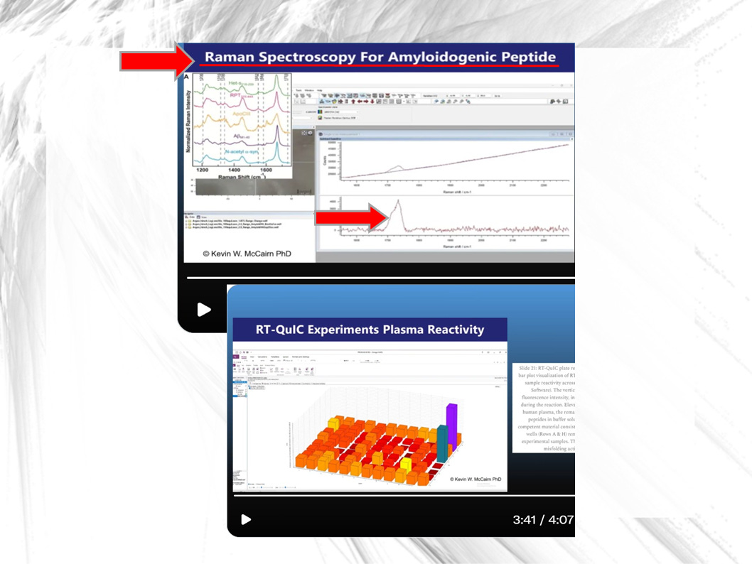
RT-QuIC (Real Time Quaking Induced Conversion): This test was conducted for Greg Harrison’s team by Dr. Kevin McCairn in Japan. It measures whether a material can seed or spread protein misfolding.
The clot samples, sent to Dr. McCairn by embalmer Richard Hirschman, tested positive. Which means that white clots can trigger misfolding in fresh blood plasma even after having been out of a body for months.
Below you see pictures of the clot samples. Click on the link below the slide to hear Dr. Kevin McCairn talk about his findings and their ramifications:
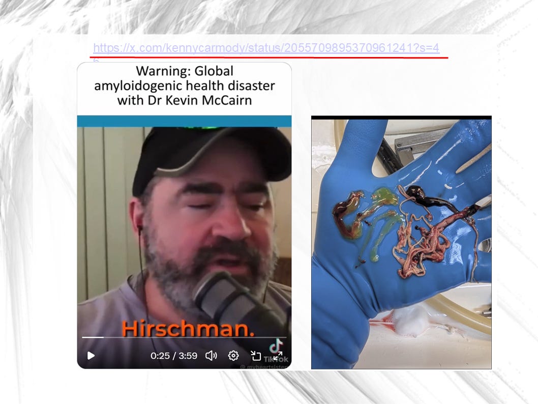
To listen to an excerpt of the interview: https://x.com/KennyCarmody/status/2055709895370961241?s=20
Harrison told me the following about the importance of McCairn’s contributions:
“Dr. Kevin McCairn’s work was the most important piece that drove us towards reconfirming his work and doing some deeper analysis.
“After we discovered the Thioflavine fluorescence, Richard Hirschman and I contacted Dr. McCairn in Japan and we convinced him to analyze twelve samples of Richard’s white clots that he had accumulated from donors ranging from the age of 33 up to 94. That’s 12 samples.
“And Kevin McCairn performed some far more sophisticated analysis that was outside of my capability and access…
“And we have to always give him due credit for being the very first one to look at these [white clots] in depth. And this work is stellar.”
The White Clots’ Seeding Capability in the Bloodstream
Further up I already mentioned the white clots’ scary seeding capability or property: McCairn took a clot sample that had been outside a human body for three months and he found that the clot was still able to cause misfolding in fresh blood plasma when brought into contact with it.
This is typical amyloid prion-like behavior: it’s not a true prion which affects the brain but it has a similar seeding effect to a prion except that it is occurring within the bloodstream.
Harrison emphasized during the interview that the research team thankfully did not find evidence that the clots are able to trigger a full prion disease (like Alzheimer’s); they believe the effect occurs in the blood only, not outside the body.
Harrison’s Summary Characterization of White Clots
Below is a slide where Harrison summarizes the characteristics of the white clots found after the mRNA Covid mass injection campaign began.
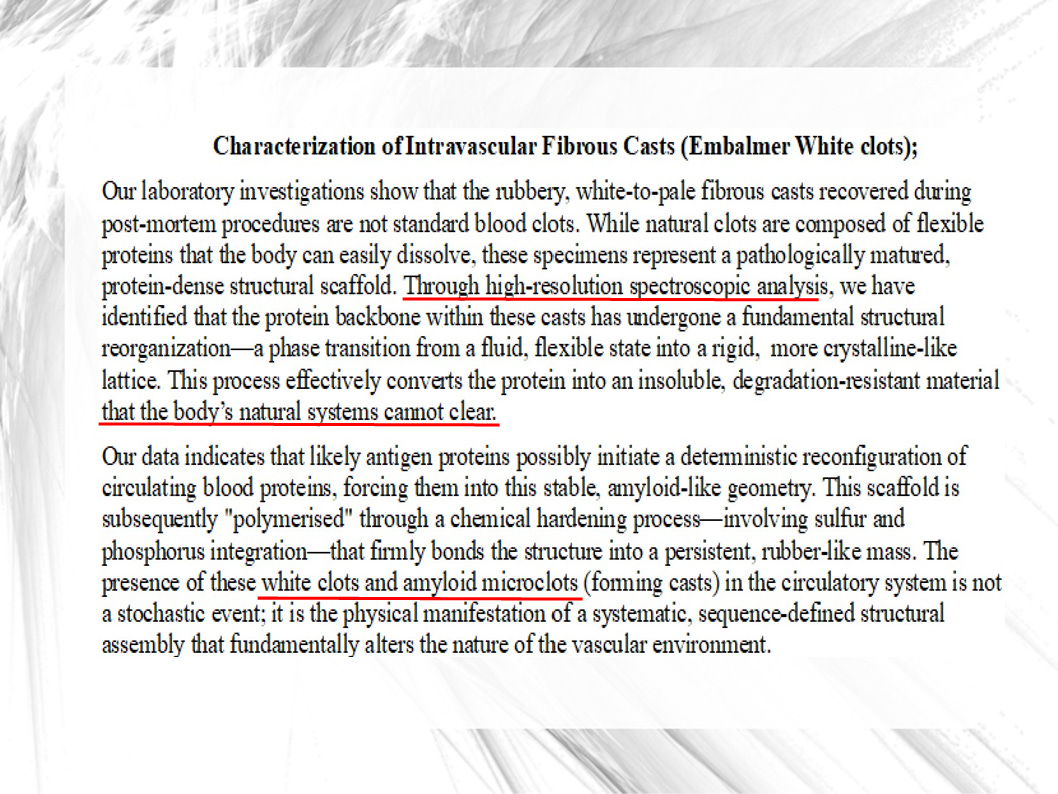
In everyday language, Harrison makes three important points here:
These white, rubbery clots are not normal blood clots.
Normal clots are made of flexible proteins that the body can eventually break down, but these specimens look like a dense, hardened protein structure instead.The proteins inside them seem to have “locked” into a rigid, crystal‑like form.
High‑resolution spectroscopy shows the clot proteins have changed from a soft, flexible state into a stiff, more crystalline, amyloid‑like lattice that the body’s cleanup systems cannot remove.Trigger proteins and chemical changes turned these into persistent, rubber‑like casts in blood vessels.
Certain antigens push circulating proteins into this abnormal shape. Chemical hardening, which involves sulfur and phosphorus, then makes the structure permanent, leaving white clots and tiny amyloid plugs that systematically change how blood vessels work.
Not Your Usual Blood Clot: White Clots Are Self‑building, Highly Stable Protein Scaffolds
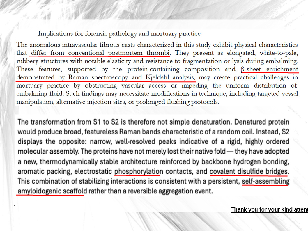
On the slide below, Harrison continues to describe the characteristics of the white clots and, in particular, the implications for embalmers and forensic pathologists.
These clots, or casts (since they resemble casts made from the vascular system), are long, pale, stretchy, and hard to break apart during embalming. Because the clots can block blood vessels, they may stop embalming fluid from flowing properly, meaning embalmers might need new techniques, such as targeting specific vessels or flushing for longer.
Tests that measure their protein structure (including Raman spectroscopy and chemical analysis) show a high amount of stiff, sheet‑like protein—a pattern typical of amyloid‑type material.
Detailed measurements of their protein signals show sharp, well‑defined peaks, which tell us the proteins have not simply fallen apart. Instead, they seem to have re‑arranged into a tight, stable, locked‑in structure held together by several kinds of strong chemical bonds, including added phosphate groups and disulfide bridges.
This suggests, Harrison writes, that what we are dealing with here are self‑building, highly stable protein scaffolds - and not a loose, temporary clump of protein.
Possible Treatments
Are there any treatments for white clots? Recently, there has been a hopeful development: not even a year ago Dr. McCairn in Japan pioneered filtering the blood of patients to physically remove clots. This treatment is called Apheresis (blood filtering).
So far only very few patients travelled to Japan to have their blood filtered. But the approximately 20 patients who have been treated are apparently showing significant improvement, with no recurrence reported at 6–8 months follow-up.
Dr. Joachim Gerlach in Germany is also working on a treatment combination showing promise for dissolving amyloid microclots.
The Paper & Next Steps
Greg Harrison’s research team is currently preparing to submit a peer-reviewed paper confirming the amyloidogenic nature of the white clots. Its publication, Greg told me, is imminent. Once it’s published, I will add the link to the Resources section below (UPDATE: The article was published in the International Journal of Innovative Research in Medical Science just one day after I released the interview with Greg. Here is the link: Raman spectroscopic Characterization of Anomalous Intravascular Fibrous Casts: Evidence for Stage-Dependent β-sheet Enriched Protein Maturation).
Greg’s team is also working on a second paper where they investigate whether spike protein is directly present in the clots. For this they use an even more targeted Raman analysis as well as what’s called NMR: nuclear magnetic resonance spectroscopy.
All of the team’s research has been entirely self-funded with no government support whatsoever.
Anyone Surprised that Health Authorities Worldwide Have No Interest in Investigating White Clots?
Well, at this point there are probably few people who are surprised that neither the Australian health authorities nor the American or European ones or any other country’s have shown the slightest interest in the white clots.
After all, why would those with blood on their hands be interested in digging their own graves?
When I asked Greg - who’s Australian - about the reaction of the Australia’s public health agencies, he told me:
“There’s no interest here whatsoever. We’re all branded conspiracy theorists and just ignored basically. So that’s why we’ve just continued on to do the work quietly.
“And we know that people like Dr. John Campbell and Doctor Philip McMillan are waiting for peer reviewed papers and probably for published reports in order to expose or should I say, alert the public to what’s going on.
“So we’re hoping that by now giving them a paper that is proposed for peer review with all the data, that they will also now take the opportunity to do videos and expose what we’ve found.
“But it’s taken a year. Yeah, we had to find a lab, we had to fund it ourselves. There’s no public funding, it’s all out of our own pocket.”
CHAPTERS
00:05 Opening And Recap Of The White Clot Mystery
00:49 Greg Harrison’s Claim: White Clots Are Amyloid‑Like
03:04 What Is Amyloidosis? From Rare Brain Disease To Blood‑Borne Systemic Form
05:50 Known Amyloid Diseases And Why Misfolding Matters
06:17 Early Analytical Work: Phosphorylation, Sulfation And Misfolded Fibrinogen
10:30 Thioflavin T Fluorescence And Elemental Signals In Embalmer Clots
14:51 Kevin McCairn’s Findings: Misfolded Amyloidogenic Fibrin And Systemic Risks
19:05 Infective Peptide Concept: White Clots Seeding Misfolding In Fresh Plasma
20:19 Microscopy Of “Calamari” Clots And RT‑QuIC Seeding Experiments
25:18 Amyloid‑Prion‑Like Behavior And Caution About True Prion Claims
27:00 Why Raman Spectroscopy Is Definitive For Detecting Amyloid Structure
28:37 Raman Fingerprints Of Clot Samples Showing Progression Toward Beta Sheets
31:18 Reference Spectra: Healthy Plasma, Fibrinogen And Spike‑Protein Bands
34:16 European Raman Lab Work And Peer‑Reviewed Paper Preparation
35:09 “Amyloidogenic Scaffolds”: Converging Raman And ThT Evidence
37:30 Novel Intravascular Scaffolds And Why They Differ From Standard Clots
38:34 Structural Reorganization: From Flexible Proteins To Rigid Crystalline Lattice
41:02 Lab Conclusion: Self‑Assembling Amyloid Scaffold, Not Reversible Clotting
42:36 Data, University Backing, And Anticipated Pushback From Institutions
43:45 Explaining Aromatic Packing And Why These Scaffolds Resist Breakdown
44:39 Possible Dissolution: Stem‑Cell Approaches And Other Anti‑Amyloid Strategies
46:09 Microclots, Strokes And Links To Long COVID And Fibrin(oid) Deposits
47:22 Microclots In Vaccinated And Unvaccinated: Role Of Open Spike Conformation
49:34 Speed Of Clot Emergence After mRNA Rollout And Survey Data
50:06 Apheresis In Japan: Blood‑Filtering To Remove Amyloid Microclots
51:08 Follow‑Up Outcomes: Patients Remaining Clot‑Free For 6–8 Months
51:29 Self‑Amplifying RNA, Broader mRNA Platform Concerns And Phosphorylation
54:02 Lack Of Interest From Australian Public Health And Self‑Funded Research
55:28 Future Tests: Focused Raman On Spike Region And Phosphorus NMR
57:31 Anticipated Impact Of Publication And How Critics Might Respond
58:02 Closing Thanks And Framing The Work As Groundbreaking
RESOURCES
Raman spectroscopic Characterization of Anomalous Intravascular Fibrous Casts: Evidence for Stage-Dependent β-sheet Enriched Protein Maturation, International Journal of Innovative Research in Medical Science; Authors: Daniel Santiago, Greg Harrison, Miklos Veres, Mark File and Dennis Planner
Australian Amyloidosis Network: https://aan.org.au/patients-and-carers/what-is-amyloidosis/
Douglas B. Kell and Etheresia Pretorius: Proteomic Evidence for Amyloidogenic Cross-Seeding in Fibrinaloid Microclots
Dr. Kevin McCairn interview: https://x.com/kennycarmody/status/2055709895370961241?s=46
My first interview with Greg Harrison from spring 2025: https://flashlightsproductions.substack.com/p/do-mrna-covid-shots-cause-the-misfolding

